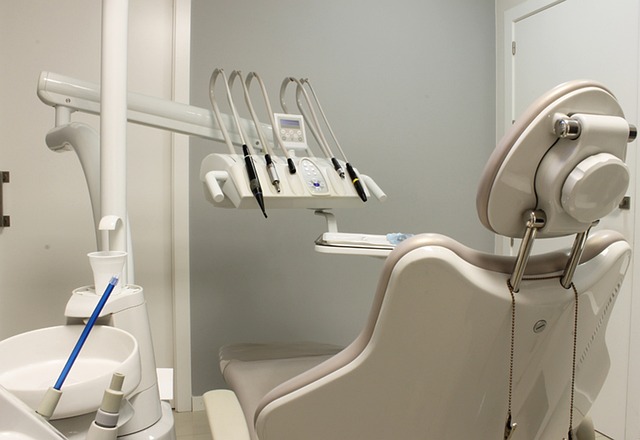
In Cambridge, dental implant surgery leverages advanced diagnostic imaging, including X-rays and CT scans, for precise treatment and optimal outcomes. These imaging tools provide detailed visualizations of the jawbone and surrounding structures, essential for assessing bone density and quality to ensure successful implant placement. The comprehensive images from these scans allow dental professionals to customize each implant to fit a patient's unique anatomy, which is crucial for stability and endurance post-surgery. During the procedure, imaging guidance helps position the implants at ideal angles and depths, critical for both function and aesthetics. The accuracy of these methods minimizes risks and complications, such as nerve or sinus injury. Patients can expect their surgery to be conducted with state-of-the-art precision using the latest in diagnostic dentistry. The detailed preoperative evaluations and post-surgical monitoring, facilitated by high-resolution imaging, ensure informed treatment decisions, long-term success, and minimal complications associated with dental implant surgery in Cambridge. This approach not only focuses on functionality and aesthetics but also prioritizes patient comfort and the longevity of the prosthetic, making it a reliable and effective solution for those with missing teeth in the region.
When it comes to dental implant surgery, precision and thorough planning are paramount. In Cambridge, advanced diagnostic imaging plays a pivotal role in ensuring the success of this procedure. This article delves into the critical importance of X-rays and CT scans in the entire process, from initial assessment to postoperative monitoring. We explore how these imaging techniques provide dentists with a detailed understanding of the patient’s oral condition, enabling tailored treatment planning for dental implant surgery in Cambridge. Join us as we uncover the benefits of integrating cutting-edge X-ray and CT scan procedures into dental implant practices, enhancing the precision and effectiveness of the treatment.
- Understanding Dental Implant Surgery in Cambridge: The Role of X-rays and CT Scans
- The Preoperative Assessment: How X-rays and CT Scans Inform Treatment Planning for Dental Implants
- The Precision of Imaging Techniques in Dental Implant Surgery: Benefits of Advanced X-ray and CT Scan Procedures
- Postoperative Monitoring: Utilizing X-rays and CT Scans to Ensure Successful Healing and Osseointegration in Dental Implant Patients
Understanding Dental Implant Surgery in Cambridge: The Role of X-rays and CT Scans

When considering dental implant surgery in Cambridge, the integration of advanced diagnostic imaging is paramount to ensure successful outcomes. X-rays and CT scans play a pivotal role in this process by providing detailed visualizations of the jawbone and surrounding structures. These images enable dental professionals to assess the bone density and quality, which are critical factors for implant placement. X-rays, particularly panoramic X-rays, offer a broad view of the entire jaw, while CT scans deliver high-resolution, three-dimensional representations that facilitate precise planning. This meticulous preoperative evaluation is essential for tailoring the implant size and position to the unique anatomy of each patient, thereby increasing the likelihood of a secure and lasting integration with the jawbone.
During dental implant surgery in Cambridge, the use of X-rays and CT scans extends beyond diagnosis. These imaging techniques guide the oral surgeon throughout the procedure, ensuring that the implant is placed at the optimal angle and depth for functional and aesthetic success. The precision afforded by these advanced imaging tools helps to minimize surgical risks and complications, such as damage to surrounding nerves or sinuses. Consequently, patients can trust that their dental implant surgery will be performed with the utmost care and attention to detail, utilizing the most accurate and effective diagnostic methods available in the field of dentistry.
The Preoperative Assessment: How X-rays and CT Scans Inform Treatment Planning for Dental Implants

Prior to dental implant surgery in Cambridge, a comprehensive preoperative assessment is pivotal to ensure successful treatment outcomes. This process begins with diagnostic imaging, where X-rays and CT scans play a crucial role in visualizing the patient’s oral cavity and surrounding structures. X-rays provide a two-dimensional image that helps identify the position of teeth, the presence of any infections, and assesses bone density—key factors for dental implant placement. CT scans offer a more detailed three-dimensional view, allowing clinicians at specialized dental implant centers in Cambridge to evaluate the anatomical relationships with nerve canals, sinuses, and critical structures, which is essential for planning the precise angle and positioning of the implants. These advanced imaging techniques enable the surgical team to tailor the implant surgery to the individual patient’s needs, optimizing both the functional and aesthetic outcomes of the procedure.
The integration of X-rays and CT scans in the preoperative assessment is a cornerstone of modern dental implantology. The high-resolution images obtained from these scans facilitate meticulous treatment planning by providing detailed insights into the patient’s oral health status, the condition of the jawbone, and the available space for the implants. In Cambridge, where dental implant surgery is highly sought after due to its effectiveness in restoring dental function and aesthetics, the preoperative imaging process is a critical step that informs the surgical strategy and enhances patient care. The information derived from these scans ensures that each implant is placed with the utmost precision, which is vital for long-term success and minimizes the risk of complications post-surgery.
The Precision of Imaging Techniques in Dental Implant Surgery: Benefits of Advanced X-ray and CT Scan Procedures

In the field of dental implant surgery, precision is paramount to ensure successful outcomes and long-lasting functionality. Advanced imaging techniques play a crucial role in this process, offering an unparalleled level of detail that traditional methods cannot match. Dental professionals in Cambridge utilise both X-rays and CT scans to accurately assess the condition of the jawbone, determine the exact positioning for implant placement, and evaluate the surrounding structures. These imaging modalities provide a comprehensive analysis of the patient’s oral anatomy, allowing for personalised treatment plans tailored to individual needs. The high-resolution images obtained from X-rays and CT scans enable dentists to identify potential complications early on, thus optimising the success rate of dental implant surgery in Cambridge. Moreover, the accuracy of these imaging techniques ensures that the implants are positioned correctly, which is essential for the integration with the bone and the longevity of the prosthetic. This meticulous approach not only enhances patient comfort but also contributes to the aesthetics and functionality of the final restoration, making dental implant surgery a reliable and effective solution for those with missing teeth in Cambridge.
Postoperative Monitoring: Utilizing X-rays and CT Scans to Ensure Successful Healing and Osseointegration in Dental Implant Patients

Following dental implant surgery in Cambridge, postoperative monitoring plays a pivotal role in the successful integration and healing of the implant. Advanced diagnostic imaging techniques such as X-rays and CT scans are instrumental in this process. These imaging modalities enable clinicians to visualize the osseointegration process, ensuring that the titanium implant is securely anchored within the jawbone. The resolution and precision of CT scans are particularly advantageous, as they provide a detailed three-dimensional view of the bone structure and implant positioning. This allows for precise monitoring of the healing progress, identifying any complications such as peri-implantitis or osseointegration failure early on. X-rays, on the other hand, are valuable for routine follow-up visits, tracking the integration over time, and detecting any shifts in the position of the implant that may occur post-surgery. The timely application of these imaging techniques is crucial for adjusting treatment plans accordingly, thereby optimizing patient outcomes and enhancing the longevity and functionality of dental implants in Cambridge patients. Regular monitoring with X-rays and CT scans not only facilitates early intervention but also contributes to the overall success of dental implant surgery.
In conclusion, dental implant surgery in Cambridge is a meticulous procedure that hinges on the precision and detail afforded by X-rays and CT scans. These imaging techniques are indispensable, from the initial diagnosis to the final stages of healing. They play a pivotal role in preoperative planning, allowing for tailored treatment strategies that optimize patient outcomes. Postoperatively, regular imaging ensures close monitoring of the integration process, guaranteeing the highest standards of care and leading to predictable and successful results. The advanced imaging modalities not only facilitate surgical precision but also underscore the commitment to excellence in dental implant surgery within Cambridge.